The human heat is a muscle, responsible for pumping blood through out the body. It begins to pump while we are still in our mothers womb and does not stop until we die. At resting, the heart will beat an average 60 beats per minute. However when under stress, this can increase to 200 beats per minute.
Unlike normal muscles the heart is made of cardiac muscle. The reason for this is its work rate. Beating over 35 million times per year or 100,000 per day,without cramping up like normal skeletal muscle it would cramp up.
Before we look at how the heart works, lets first look at the structure of the heart.
The first thing you’ll notice is that it looks nothing like the cartoon drawings. Its roughly the size of your fist and sits more so on the left side of your body, protected by your lungs and rib cage.
The heart has 4 chambers. A right atrium (top), right ventricle (bottom), left atrium (top) and a left ventricle (bottom).
These chambers are essentially rooms in the heart. The blood fills up and then is released into the next room (chamber). The important thing to note about each chamber is the thickness of the walls. The left ventricle is the largest and strongest chamber.
Separating each room is a door. These doors are called valves, and there are 4 of them.
The tricuspid valve regulates blood flow between the right atrium and right ventricle.
The Pulmonary valve controls blood flow from the right ventricle to the pulmonary arteries.
The mitral valve lets blood from your left atrium to the left ventricle
Finally the aortic valve releases blood from the left ventricle to the aorta.
So how does the blood get into and out of the heart?
The VenaCava (Vein) brings de-oxygenated blood to the right atrium.
The Pulmonaryartery (Artery) delivers blood from the right ventricle and to lungs.
The Pulmonaryvein (Vein) brings oxygenated blood from the lungs to the left atrium
Finally, the Aorta (Artery) delivers the oxygenated blood to the whole body.
Above is an echocardiogram of a 40 year old male.
Notice the flaps opening and closing
So as stated in the above video:
Blood is returned from the body through the superior and inferior vena cava
Blood enters the heart in the right atrium
Blood the passes from the right atrium to the right ventricle
The blood is then pumped through the pulmonary artery to the lungs
Blood releases its carbon dioxide and gains oxygen. It becomes re-oxygenated.
The blood then leaves the lungs and re-enters the heart through the pulmonary vein
The blood in poured into the Left atrium
The blood then enters the left ventricle
The blood is squeezed out of the ventricle and passed through the Aorta to the body.
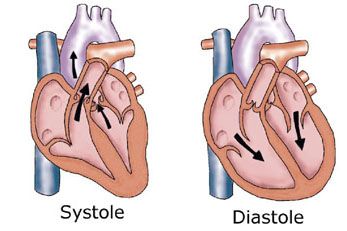
The heart has two sides to it. One is oxygenated, while the other is de-oxygenated. This is referred to as the double pump system. The septum separates the oxygenated from the non-oxygenated blood. The beating heart goes through a cycle of contraction and relaxation. Contraction is known as systole while relaxation is known as diastole.
To left is an MRI scan of an individuals heart, from different angles
During systole the ventricles contract forcing blood into the body and lungs. The right ventricle contracts a little before the left ventricle.
When blood leaves the heart it goes through the Aorta, this artery is thick as the blood is under high pressure. This is because the blood has to go to the entire body.
During diastole the ventricles relax and are filled with blood coming form the atria.
Click here for a heart dissection
Double pump system
During systole, the Tricuspid and mitral valve slam shut. This causes the ‘Lub’ sound or 1st hear sound.
During diastole, the pulmonary and aortic valves shut. This causes the ‘Dub’ sound or 2nd heart sound.


 Structure
How it flows
Structure
How it flows