


The Trachea is a long tube which travels from the pharynx to the lungs.
Its held open with ‘C’ shaped rings of cartilage. This prevents the trachea from collapsing when there is no air flow.

Unlike the respiratory system of fish, our lungs are inside us. The reason that we have our lungs inside is so they remain moist. The moister allows oxygen and carbon dioxide to easily diffuse across into the lungs.
Lets discuss the structural components of the respiratory system
The first thing you will notice is that both the nose and mouth link to the Trachea.
This means air taken in through the mouth goes to the same place as air taken through the nose, however it is the preferable that air is taken in through the nose. This is because air can be filtered, moistened and warmed through the nasal passages.
Air taken in is pushed to the back of the throat (pharynx).

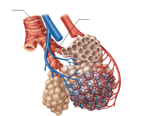
Moving down the trachea, it eventually splits forming two tubes called Bronchi.
Within the Trachea and Bronchi are ciliated cells (click here for information on cilia) which produce mucus.
The mucus traps dust and bacteria brought in during breathing. The cilia then push them back up towards the mouth, so it can be swallowed.
The Bronchi continue to branch into smaller tubes called Bronchioles.
They eventually lead into small air sacs called Alveoli. This is where the majority of gas diffusion occurs. Our lungs contain over 300 million alveoli.
Capillaries are wrapped around the Alveoli. This allows diffusion of gasses through the blood stream. We will talk more about this later on.
By constantly branching out we increase the SA:V ratio. Our lungs have roughly the same SA of a tennis court.
If we have a look at the lungs we can see a difference between the right and left.
The right lung has 3 lobes, where as the left only has 2 lobes. Another thing you will notice is dip in the left lung.
This is called the Cardiac notch. The heart sits just behind here.
Wrapped around the lungs is are the ribs. The ribs expand upwards and outwards while inhaling and contract downwards while exhaling.
The Diaphragm is another extremely important muscle in breathing. It is a thin sheet of muscle and tendon which separates the abdominal cavity from the chest cavity.
As we inhale, the Diaphragm contracts pulling air in. As the Diaphragm relaxes it pushes air out of the lungs.
This is shown by the yellow line in the animation to the left.
Now that you know the basic components of the human respiratory system. Lets take a look how oxygen gets from your lungs to all your body cells.
Click here to see a pluck dissection
As you can see from the video to the left, Your vocal cords allow you to change your pitch without inhibiting air flow.

At the top of trachea is the Larynx (voice box). This is where the vocal cords are located.
The vocal cords allow us to change our pitch and tone. When we whisper, yell, talk in a deep voice or high pitch voice, we use our vocal cords.

