The nerve cell is probably one of the most complex and yet most important cells in your body. In order to understand how the nerve cell works in your body, you first have to learn its basic structure. Once you can accurately identify all the parts of a neuron, then we can begin to discuss how they function within the body.
Structure
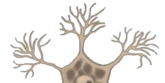
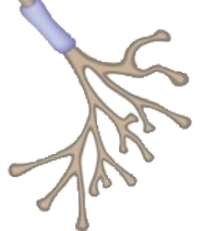
Observe the image to the right. This is the basic structure of a nerve cell. Scroll over each word to show its location.
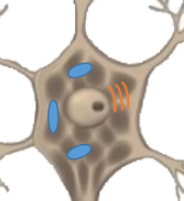
These are short projections from the cell body. They receive and carry nerve impulses towards the cell body
Dendrites
This contains the nucleus, mitochondria and other organelles which keep the cell functioning.
The nucleus is the storage centre for DNA.
Cell body/ soma and nucleus
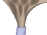
The myelin sheath is an extremely important part of all nerve cells. This fatty tissue speeds up transmissions between nerve cells and prevents the signal from jumping to adjacent nerve cells. In infants myelin is not fully developed, which is why they have poor co-ordination.
A nice analogy of myelin is to compare it to the insulation on a wire. When wires is not insulated their signal can jump to other wires.
The Schwann cells are responsible for the production of myelin
These are the space between each myelinated part of the axon. Action potential takes place here.
Node of Ranvier
This is the end of the neuron. Neurotransmitters are released from this point and attach to receptors on muscles, glands or other neurons.
Axon endings/ Axon terminals
As you can imagine the nervous system is extremely large and complex, thus requiring multiple different types of neurons. We can classify nerve cells by, shape, size or function. We will focus on function. There are three main types of neurons:
Motor neurons are attached to muscle or gland. In the animation to the right, the neuron receives a signal and sends a message to a muscle, causing it to contract.
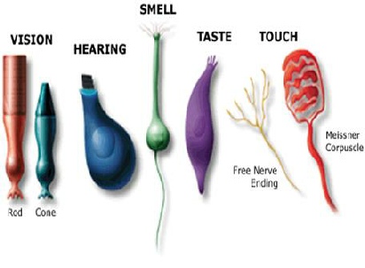
Sensory neurons are more complex. Observe the animation to the right. This is a sensory neuron located on the skin, to detect touch.You should notice a few key differences:
There are no dendrites at the top. Instead they have receptors.
The nucleus is located away from the receptors and dose not have dendrites protruding from it.
Their function is to detect a change and report it. Sensory neurons can be broken up into many different categories depending on what the detect.
Inter neurons are similar in structure to motor neurons, however they do not attach to a muscle, instead they send their signal to a motor neuron or another inter neuron. You can see in the animation to the right that as the sensory neurons are stimulated the inter neurons carry the message to the spine.